
Figure 1.
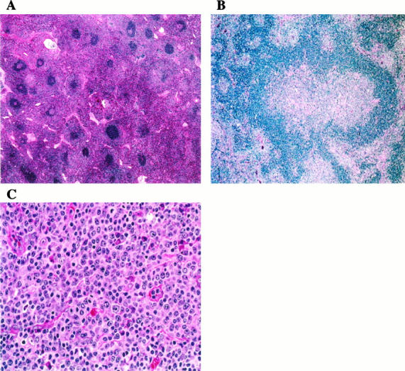
Histopathology of lymph nodes. A: The paracortex is markedly expanded by lymphocytes with pale pink cytoplasm. Primary and secondary follicles are numerous. Some follicles show regressive changes (upper right ). H&E; original magnification, ×100. B: Progressive transformation of germinal centers was a focal but relatively frequent finding. H&E; original magnification, ×200. C: The paracortex is populated by lymphocytes, plasma cells, and immunoblasts. Note frequent mitotic figures. H&E; original magnification, ×600.