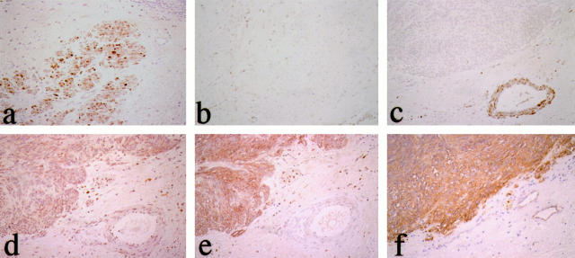
Figure 1.
Immunohistochemical evaluation of myosin heavy chain isoforms in GIST. Smooth muscle cells (SMCs) of muscular layer and blood vessels of the stomach are positive for SM1 and SM2 (a) but negative for SMemb (b). The neoplastic cells of GIST are completely negative for SM1 and SM2 (c). On the other hand, the neoplastic cells show diffusely positive staining for SMemb (d), KIT (e), and CD34 (f). ABC immunostaining; magnification, ×30.

