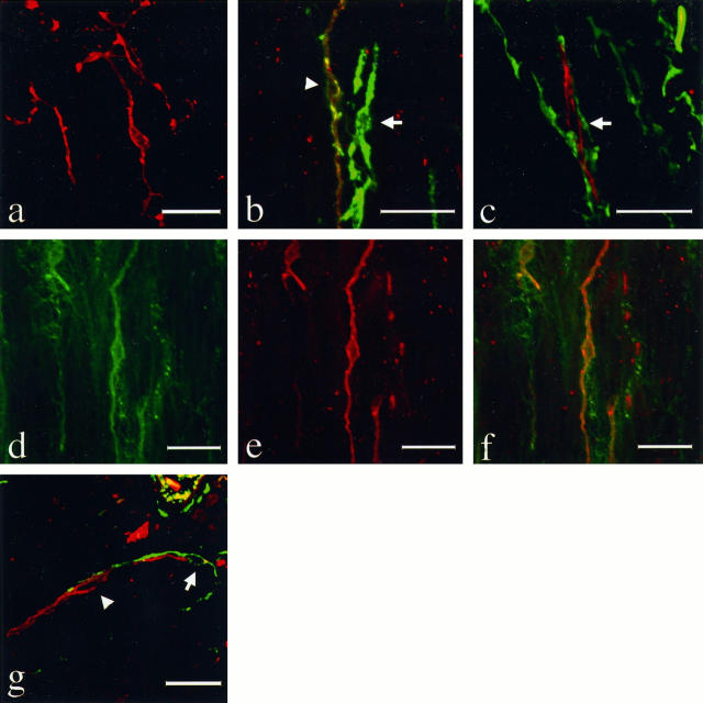
Figure 2.
Fluorescence double immunolabeling of the interstitial cells of Cajal (ICCs) of the stomach (a to f) and small intestine (g). Immunohistochemistry was applied to 15-μm-thick sections, which had been treated by the AMeX method, using a combination of polyclonal anti-KIT antibody with monoclonal antibodies to other substances. TRITC-conjugated swine anti-rabbit IgG and FITC-conjugated goat anti-mouse IgG were used for visualization of the immunoreaction, respectively. The sections were examined with a confocal laser scanning microscope with an excitation wavelength of 568 nm for TRITC (red) and 488 nm for FITC (green). KIT-positive ICCs (red) in the muscular layer form a complex cell-network (a, magnification, ×625). KIT-immunoreactive cells are in most cases positive with CD34 (b, left, arrowhead, magnification, ×800), but there is also a KIT-negative, CD34-positive ICCs-like cell (b, right, arrow) and KIT-positive and CD34-negative ICCs-like cells (c, arrow, magnification, ×800). On the other hand, SMemb (d) and KIT (e) immunoreactivities are observed in nearly all of the same cells (f, magnification, ×575). In the muscular layer of small intestine, most of the KIT-positive cells were negative for CD34 (g, arrowhead) but were present in the vicinity of KIT(−) CD34(+) ICCs-like cells (g, arrow, magnification, ×600). The latter type of interstitial cells were more frequent and distributed more widely in the tunica muscularis and subserosa. Bars, 20 μm.

