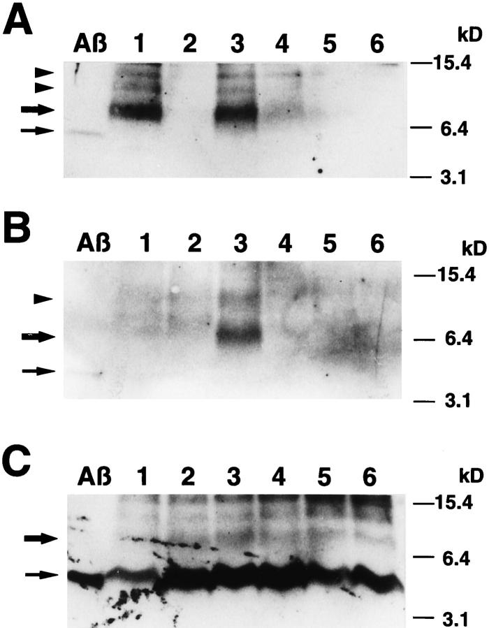
Figure 3.
Western blots of representative EIA-negative specimens and PDAPP transgenic mouse brains. A, B: Representative cases showing the presence of SDS-stables Aβ42 (A) and Aβ40 (B) dimers. Each left-most lane is loaded 10 pg of synthetic Aβ1–42 (A) or Aβ1–40 (B). An upper arrowhead in A or an arrowhead in B indicate a 12-kd band, presumably representing Aβ trimer. A lower arrowhead in A indicates an ∼8-kd band, perhaps representing anomalously folded Aβ42 dimer. 36 Small and large arrows in A and B indicate Aβ monomer and Aβ dimer, respectively. C: The effects of postmortem delay on the molecular form of Aβ42 in the transgenic mice. The mice were kept at room temperature for 0 (lane 1), 2 (lane 2), 4 (lane 3), 6 (lane 4), 12 (lane 5), and 18 (lane 6; see text) hours after death and processed for Western blotting with BC05. No immunoreactivity with BA27 was detected on the blot (data not shown). The leftmost lane is loaded 10 pg of synthetic Aβ1–42. Small and large arrows indicate Aβ42 monomer and Aβ42 dimer, respectively.

