
Figure 4.
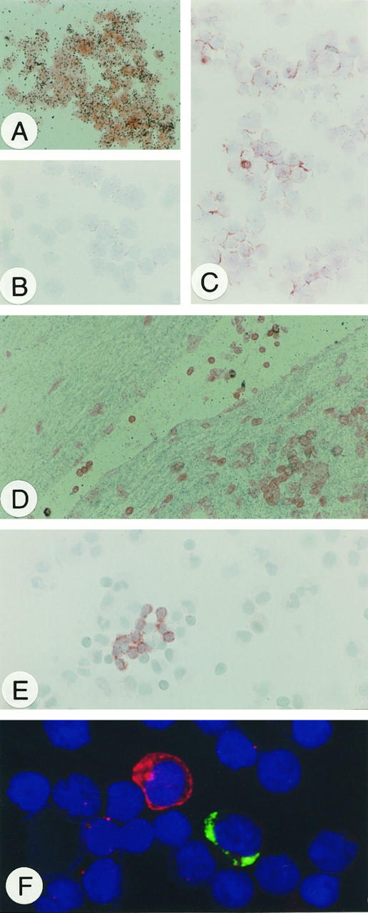
A: Hybridization of CD95L mRNA in PMA/ionomycin-stimulated Jurkat cells. B: Immunohistochemical detection of CD95L protein in unstimulated, C: PMA/ionomycin-treated Jurkat cells. D: In situ hybridization of CD95L mRNA in activated T cells within blood vessels. E: CD95L protein detection by immunohistochemistry in activated T cells in inflammatory pleural effusions. F: Immunofluorescence double staining in lymphoid cells from a cystic lymphangioma. CD95L (red) is detected cytoplasmatically and on the surface of a single lymphoid (probably activated T) cell that lacks IgG expression (green). Original magnification, ×100 (A to E); ×400 (F).