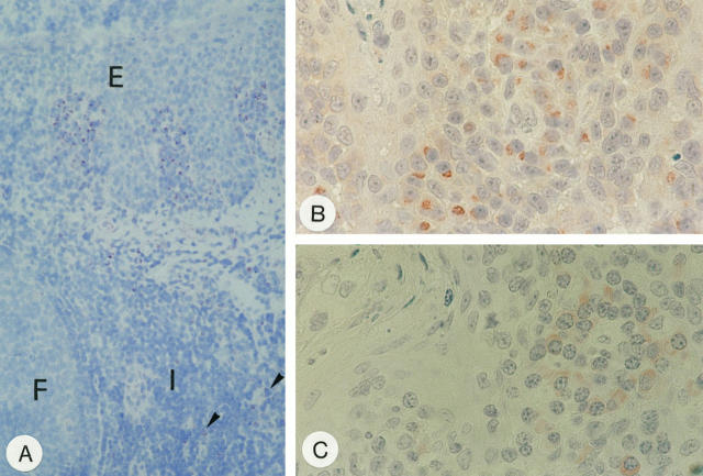
Figure 5.
CD95L protein detection in tissue. A: Immunohistochemistry with mAb G247–4 on frozen section of the tonsil. Note groups of stained cells beneath the squamous epithelium (E) of the crypts with only a few weakly stained cells scattered in the interfollicular areas (I) (arrow heads). F, lymphoid follicle. Immunohistochemistry on paraffin sections using mAb G247–4 (B) and 139 (C). Note different subcellular staining patterns yielded by the two mAbs. In B, nuclear morphology of plasma cells is artificially altered because of microwave irradiation. Plasma cells, however, are readily identified by their broad cytoplasmic rim around an eccentrically located nucleus. Original magnification: ×50 (A); ×157 B and C.

