
Figure 6.
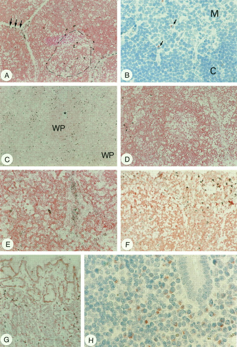
CD95L detection in lymphoid organs. A: In situ hybridization showing CD95L+ cells at the corticomedullary boundary (broken line) and in the subcapsular cortex (arrows) of the thymus. B: Immunohistochemistry reveals weakly positive cells only at the corticomedullary boundary (arrows; M, thymic medulla; C, cortex). C: Detection of CD95L mRNA in the spleen by in situ hybridization. Positive cells are scattered in the red pulp, whereas the white pulp (WP, an arteriole is highlighted by the star) is largely devoid of CD95L+ cells. D: CD95L mRNA-expressing cells in the lymph node were detected by in situ hybridization mainly in the interfollicular areas/medullary cords of the lymph node. E: In some cases, high endothelial venules were labeled. F: In situ hybridization of CD95L mRNA of the tonsil; note strongly labeled cells beneath the surface epithelium. G: Gastric fundus in chronic gastritis of the stomach. Note CD95L+ cells scattered in the lamina propria with in situ hybridization. H shows numerous stained cells in the mucosa of the appendix, most of which are plasma cells. Note again that nuclear morphology of plasma cells is artificially altered by microwave irradiation as in Figure 5B ▶ . Original magnification, ×25 (A, C, D, F, and G); ×100 (B); ×50 (E); ×157 (H).