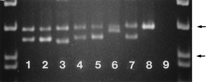
Figure 7.
Analysis of the PCR amplification products of the exon 11 of c-kit in malignant GISTs. Note the different size mutant bands in different cases. Lane 1: Case 20. Lane 2: Case 34. Lane 3: Case 32. Lane 4: Case 21. Lane 5 : Case 27. Lane 6: Case 42. Lane 7: Case 30. Lane 8: Placenta (germline), Lane 9: Negative control. The samples are flanked by PhiX174 DNA/HinfI markers (Promega, Madison, WI). Arrows indicate 200- and 151-bp fragments. 5% polyacrylamide gel electrophoresis.

