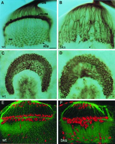
Figure 1.
brakeless is required for R-cell projections. (A and B) R-cell projections were visualized with mAb 24B10 staining. In the wild type (A), R-cell axons project from the developing eye disk through the optic stalk (os) into the optic lobe. The dense staining layer in the lamina (la) consists mainly of expanded R1-R6 growth cones. R7 and R8 axons pass through the lamina into the medulla (me), where they form a highly ordered innervation pattern. In bks mutants (B), many R1-R6 growth cones fail to stop at the lamina and migrate into the medulla instead (also see Fig. 3B), leaving many uninnervated regions in the lamina. Consequently, abnormal thicker bundles and excessive growth cones appear in the medulla. The arrowhead in B indicates the terminus of Bolwig's nerve. (C and D) Wild-type (C) and bksP1 (D) third-instar larval optic lobes stained with anti-Dachshund. Dachshund is a nuclear protein expressed in the developing lamina neurons. Lamina neurons differentiate normally in bks mutants (D). (E and F) Optic lobes stained with the RK2 antibody recognizing Repo, a nuclear protein expressed in glial cells. In the wild type (E), R1–6 growth cones are surrounded by rows of RK2-positive glial cells. Although these cells migrate correctly into the target region and differentiate in a bks4 optic lobe (F), they appear less organized than the wild type, likely because of the abnormal R-cell innervations. (Bar = 20 μm.)

