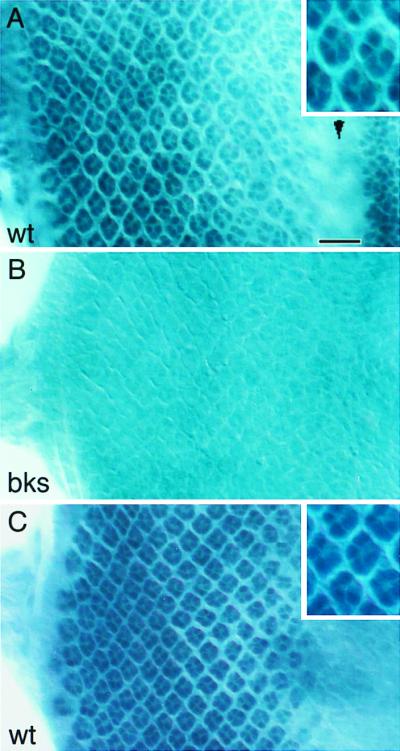
Figure 4.

Expression pattern and subcellular localization of the Brakeless protein. Third-instar larval eye discs were stained with anti-Bks antibody (A and B) or anti-Elav antibody (C). Posterior is to the left. An apical focal plane is shown. (A) In the wild type, Bks was detected in the nuclei of developing R-cells behind the morphogenetic furrow (arrowhead). The intensity of staining increases gradually from anterior to posterior. Weak Bks staining also was seen in cells located anterior to the morphogenetic furrow. The inset is a high magnification view of the staining. (B) No Bks staining was seen in bks4 mutants. (C) The nuclei of differentiating R-cells in the wild type was visualized with anti-Elav staining. The inset is a high magnification view of the nuclear staining. Note that the Bks staining pattern in the inset of A is similar to that in C. (Bar = 15 μm.)
