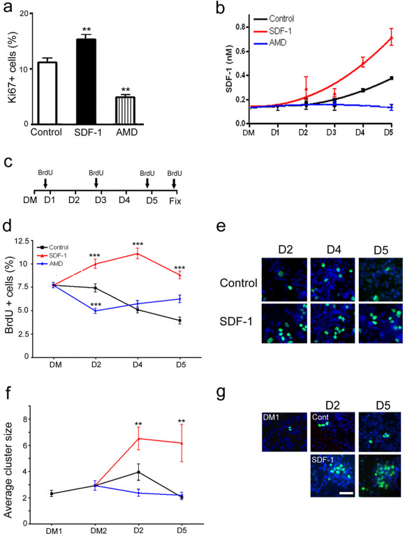
Figure 1.
SDF-1 signaling regulates cortical cell proliferation. (a) Graph illustrates a significant increase in proliferation (Ki67+) following chronic exposure to SDF-1. (b) ELISA measurements on medium collected from control and treated cortical cultures, DM – defined medium. (c) Experimental scheme for BrdU pulsing and cluster analysis; D1–D5 corresponds to 5 consecutive days of treatment with control, SDF-1 medium or AMD3100, DM corresponds to cultures in defined medium prior to the application of any treatment. Arrows indicate the time points of BrdU applications. (d, e) Graph illustrates the rate of cell proliferation as shown by the percent of BrdU+ cells at different days of treatment, while (e) shows representative images of BrdU staining (green) against counterstained cells (blue). (f, g) Graph illustrates the average BrdU cluster size at different days of treatment and (g) shows representative images of cell clusters; DM1 and DM2 correspond to cultures in defined medium for 24 hr and 48 hr, respectively. Scale bar: 100 μm.

