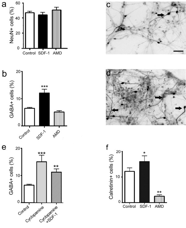
Figure 4.
SDF-1 regulates the differentiation of cortical neurons. (a) Graph shows the percent of postmitotic neurons (Neu-N + cells) in control and treated cortical cultures. (b-d) Graph illustrates the percent of GABA+ neurons in various treatments applied to cortical cultures (b). Examples of images from control (c) and SDF-1 treated (d) cultures stained for GABA immunoreactivity; arrows indicate labeled cells. (e) Treatment with cyclopamine (Shh antagonist) alone or together with SDF-1 increased the GABA expression, suggesting a sonic independent mechanism. (f) Graph shows the percent of calretinin immunoreactivity in control and treated cultures. Scale bar: 100 μm.

