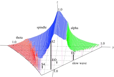
Figure 7.
Brain stability zone. The surface is shaded according to instability, as labelled (dark grey=spindle, light grey at right=alpha, light grey at left=theta), with the front right-hand face left transparent as it corresponds to a slow-wave instability. Approximate locations are shown of alert eyes-open (EO), relaxed, eyes-closed (EC), sleep-stage 2 (S2) and sleep‐stage4 (S4) states, with each state located at the top of its bar, whose x–y coordinates can be read from the grid.

