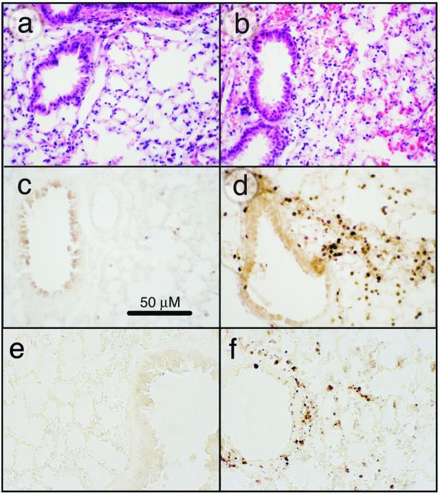
Figure 1.
Effects of ricin on lung tissues. C57BL/6 mice were instilled intratracheally with 50 μl of saline (a, c, e) or 20 μg of ricin/100 g body weight (b, d, f), and tissues were harvested at 48 hours. a and b: H&E-stained tissue sections. Tissue from ricin-instilled lung (b) showed perforations in the bronchiolar epithelium, evidence of hemorrhage, and influx of leukocytes. c and d: Immunohistochemical detection of Gr-1. Tissue from ricin-instilled lung showed accumulation of Gr-1-positive cells in peribronchiolar (d) and perivascular locations (not shown). Counterstaining of nuclei with methyl green (not shown) confirmed that the Gr-1-positive cells were neutrophils. e and f: Immunohistochemical detection of activated caspase 3. Original magnifications, ×400.

