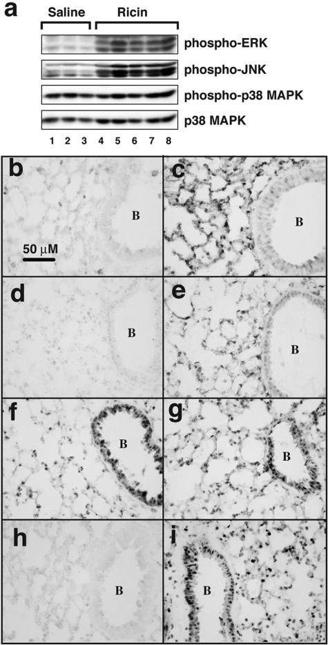
Figure 3.
Activation of signaling pathways in lung tissues. a: Protein lysates from three mice instilled with saline and five mice instilled with 20 μg of ricin/100 g body weight were harvested 48 hours later and subjected to immunoblotting using antibodies against the phosphorylated forms of ERK, JNK, and p38 MAPK and against the nonphosphorylated form of p38 MAPK (as loading control). Tissues from mice instilled with saline (b, d, f, h) or ricin (c, e, g, i) were subjected to immunohistochemical staining using antibodies against phospho-ERK (b, c), phospho-JNK (d, e), phospho-p38 (f, g), and the p65 subunit of NF-κB (h, i). B denotes the interior of a bronchiole. Original magnifications, ×400.

