Figure 1.
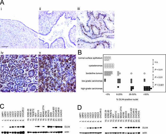
DLX4 expression in normal ovary and epithelial ovarian tumors. A: Representative examples of immunohistochemical staining of DLX4 in specimens of normal ovary (i), cystadenoma (ii), borderline tumor (iii), and low-grade (iv) and high-grade (v) ovarian carcinomas. B: Summary of DLX4 staining in clinical specimens. An average percentage of epithelial cells with DLX4-positive nuclear staining was determined for tissue cores of each case. Each symbol represents an individual case. Statistical significance of differences in percentage of DLX4-positive cells between the indicated groups of cases was calculated by the Mann-Whitney U-test. P values >0.05 were considered not significant (n.s.). C: Southern blot analysis of DLX4 RT-PCR products in specimens of borderline tumor (lanes 1 and 2), low-grade ovarian carcinoma (lanes 3 to 5), high-grade ovarian carcinoma (lanes 6 to 8), nontumorigenic ovarian surface epithelial cell lines (lanes 10 to 12), and ovarian cancer cell lines (lanes 9 and 13 to 23). D: Detection of DLX4 protein by Western blot in the same tissue specimens and cell lines analyzed by RT-PCR in C. Detection of actin RT-PCR products and protein were included as controls. Scale bar = 50 μm.

