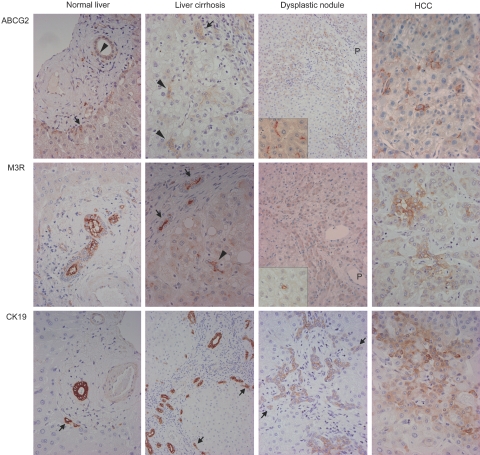
Figure 2.
Single immunostaining of ABCG2, M3R, or CK19 in normal liver, liver cirrhosis, dysplastic nodules, and HCCs. In normal liver, ABCG2, M3R, and CK19 are expressed in bile ducts (arrowhead in ABCG2) and periportal hepatocytes (arrows in ABCG2 and CK19). In liver cirrhosis, ABCG2, M3R, and CK19 expressions are observed in the bile ductules (arrows) and the canals of Hering (arrowheads). Structures indicated by arrowheads have small and inconspicuous lumen, and are not embedded in connective tissue of portal tracts, but surrounded by mature hepatocytes. These histological features suggest that these structures are canals of Hering. In dysplastic nodules, these three molecules are expressed in dysplastic hepatocytes around the portal tracts (P) (arrows in CK19). ABCG2 are mainly expressed on the canalicular membrane of hepatocytes (inset). M3R is also expressed in the small ductular structures resembling the canals of Hering within the dysplastic nodule (inset). CK19 expression is also expressed in bile ductule-like structure in intranodular portal tracts. In HCC, these three molecules are expressed in the cytoplasm or cell membranes of some cancer cells. Original magnifications: ×400 (normal liver, liver cirrhosis, HCC, and insets); ×200 (dysplastic nodule).

