
Figure 5.
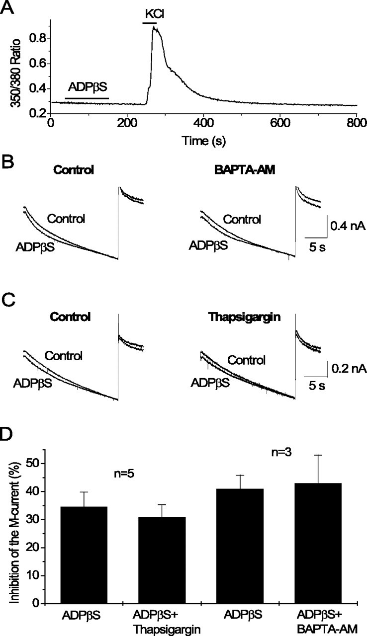
Release of intracellular Ca2+ is not involved in M-current inhibition by P2Y1 receptors in hippocampal pyramidal cells. A, Changes of intracellular Ca2+ measured as the 350/380 nm fluorescence ratio recorded at 1 Hz from a pyramidal-like neuron loaded with fura-2. Bars indicate time of drug application. Note that ADPβS at 3 μm does not induce any changes of intracellular Ca2+, whereas subsequent application of KCl at 60 mm induces a substantial increase. B, C, Currents recorded in response to voltage ramps (20 s) from −20 to −90 mV (as in Fig. 3A) in control and after application of ADPβS at 3 μm before (Control) and after 15 min superfusion of the same cell with BAPTA-AM at 10 μm (B) or with thapsigargin at 1 μm (C). D, Mean percentage inhibition of M-current by ADPβS before and after 15 min superfusion with BAPTA-AM or thapsigargin. Error bars show SEM of inhibition of the M-current by ADPβS measured at −30 mV from steady-state current–voltage relationships. n indicates the number of experiments.