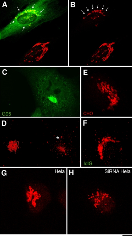
Figure 3.
The Golgi ribbon is fragmented in GM130-depleted cells. (A and B) HFs were injected with the N309 anti-GM130 antibody and after 1 h at 37°C they were fixed and stained for N309 (A; green) and giantin (A and B; red). In noninjected cells (lower right), giantin staining is reticular and continuous; in N309-injected cells (top left) it shows discrete small puncta (A and B, arrows). (C and D) COS7 cells were transfected with GFP-G95, and 20 h later (when no endogenous GM130 was detectable; Supplementary Figure S1) they were fixed; GFP-G95 fluorescence (C; green) and giantin staining (D; red) are shown. Note that the staining pattern for giantin is disorganized in transfected cells (D, asterisk; right) compared with nontransfected cells (D, left). (E and F) CHO (E) and ldlG cells (F) were stained for giantin (red). Note that the regular pattern of interconnected ring-like structures in CHO cells is absent in ldlG cells, which show small solid dispersed puncta. (G and H) Hela cells were mock-treated (G) or treated with siRNAs for GM130 (72 h; H) and fixed and stained for giantin (red). Bar, 3 μm (A, B, E, and F); 10 μm (C and D); 2 μm (G and H).

