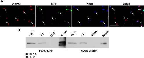
Figure 9.
Interaction of Kifc1 and Kif5B in early endocytic vesicles. (A) Texas Red ASOR-containing early endocytic vesicles from wild-type mouse liver were perfused into optical chambers and incubated with Kifc1 antibody followed by incubation with Alexa 488-labeled secondary antibody. After washing, the vesicles were incubated with antibody to Kif5B followed by Cy5-labeled secondary antibody, as described in Materials and Methods. Vesicles were visualized in the rhodamine channel, and antibody staining was visualized in the FITC (Kifc1) and Cy5 (Kif5B) channels. A typical experiment in which ASOR (red), Kifc1 (green), and Kif5B (blue) were examined is shown. Vesicles in white in the merged image (right) represent early endocytic vesicles that are associated with both motors simultaneously. Bar, 10 μm. (B) To examine whether Kifc1 and Kif5B can exist in a complex, 293T cells that endogenously express Kif5B were transfected with FLAG-Kifc1 or FLAG vector alone, and a FLAG immunoprecipitate was examined by immunoblot by using antibody to the kinesin heavy chain (KHC). Lanes in the immunoblot are cell lysate (input), representing 2% of total volume; material flowing through the anti-FLAG-agarose beads (FT), representing 2% of total volume; material in the buffer wash (wash), representing 2% of total volume; and material released from the beads after incubation in sample buffer (beads), representing 50% of bound material.

