Figure 2.
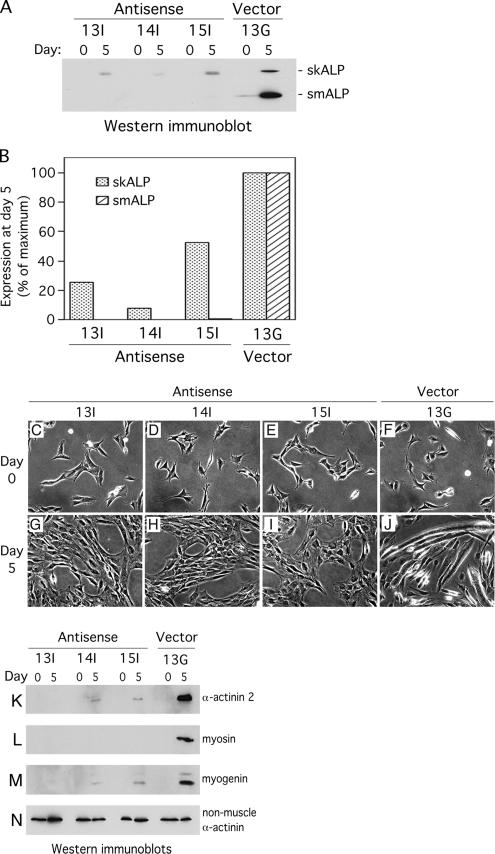
ALP-antisense C2C12 clones fail to differentiate when placed in differentiation-promoting conditions. Expression levels of smALP and skALP were analyzed in 3 ALP-antisense clones (Antisense 13I, 14I and 15I) and in 1 empty vector clone (Vector 13G). (A) smALP and skALP were detected by Western immunoblotting in proliferating cells (day 0) and in cells placed in differentiation medium for 5 d (day 5). (B) Densitometric analysis of the bands detected by Western immunoblotting at day 5 in differentiation medium. The data are expressed as a percentage of the maximum protein expression detected for the Vector 13G clone. This graph represent the densitometric analysis of the autoradiography shown in A, representative itself of five different autoradiographies. (C–J) Phase contrast pictures of the 3 ALP antisense clones (Antisense 13I, 14I and 15I) and the empty vector clone (Vector 13G). The cells were grown in proliferation medium (day 0) then placed in differentiation medium for 5 d (day 5). (K–N) Cell lysates were prepared from the 3 antisense clones (Antisense 13I, 14I and 15I) and the empty vector clone (Vector 13G) grown in proliferation medium (day 0) or placed in differentiation medium for 5 d (day 5). Expression of 3 different muscle-specific proteins was detected by Western immunoblotting: (K) α-actinin 2; (L) myosin; (M) myogenin. As a control, the nonmuscle isoform of α-actinin was detected using the same technique (N).

