Figure 5.
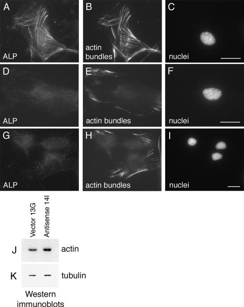
Loss of actin bundles in ALP-antisense C2C12 cells. Immunofluorescence microscopy of control Vector 13G C2C12 cells (A–C) and Antisense 14I C2C12 cells (D–F and G–I) grown in proliferation-promoting conditions. Cells were triple-labeled for ALP (A, D, and G), actin bundles (B, E, and H), and nuclei (C, F, and I). Only few short actin bundles are detected in ALP-antisense C2C12 cells. Total actin and tubulin were detected by Western immunoblotting in control C2C12 cells (Vector 13G) and ALP-antisense C2C12 cells (Antisense 14I) grown in proliferation medium. Bar, 30 μm.

