Figure 6.
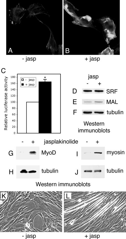
Differentiation of the ALP-antisense C2C12 cells after jasplakinolide treatment. ALP-antisense C2C12 cells were placed in differentiation-promoting conditions in absence or in presence of 0.16 μM jasplakinolide for 1 h and then grown in differentiation medium for 1 d (A–F), 3 d (G and H) or 7 d (I–L). Fluorescence microscopy was performed to visualize F-actin in ALP-antisense C2C12 cells treated (B) or not (A) with jasplakinolide. SRF activity was determined in a similar experiment where the jasplakinolide-treated cells were previously cotransfected with the SRF reporter plasmid pSRE3-Luc and a Renilla standardization reporter plasmid (C). Luciferase activities are expressed after correction for Renilla reporter activities. Results are the mean ± SEM of three independent experiments. Asterisks indicate statistical significance at p < 0.003 according to the unpaired Student's t test. Cell lysates were analyzed by immunoblotting using SRF (D), MAL (E), MyoD (G), myosin (I) and tubulin (F, H, and J) antibodies. Phase contrast images of ALP-antisense C2C12 cells treated (L) or not (K) with jasplakinolide.

