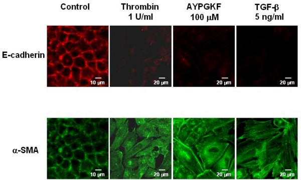
Figure 3.

Phenotypic changes in A549 cells stimulated with PAR4 agonists or TGF-β. Immunofluorescence images for a specific marker for epithelial cell (E-cadherin; rhodamine red, upper panel) or myofibroblast (α-SMA; FITC green, lower panel) captured with confocal lasar microscopy. Cells were treated with or without (control) various agonists (thrombin, AYPGKF-NH2 or TGF-β) for 72 h, and stained for E-cadherin or α-SMA using specific antibodies as described in Method section.
