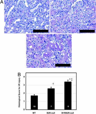
Fig. 4.
Histological changes in the postischemic kidneys of WT, B2R-null, and B1RB2R-null mice. (A) Periodic acid Schiff's staining. Shown are stainings for WT (Upper Left), B2R-null (Upper Right), and B1RB2R-null (Lower) mice. (Scale bar, 100 μm.) (B) Semiquantitative scoring for histological changes in the proximal tubule according to the criteria of Jablonski et al. (56). The sham-operated mice all a score of zero. The black columns show the scores of the ischemia groups. †, P < 0.05 vs. WT; ‡, P < 0.05 vs. B2R-null. The data are means ± standard errors, with the numbers of animals shown in white digits.

