
Figure 1. Avian vaginal morphology.
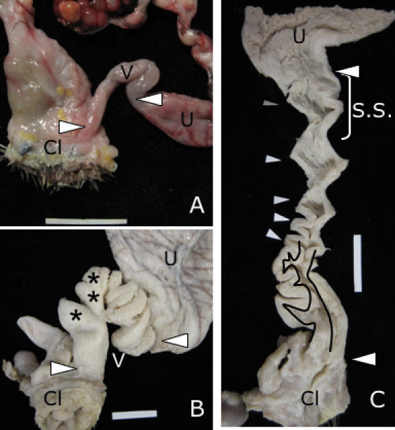
(A) Typical tubular avian vagina from domestic Pheasant (Phasianus colchicus) (connective tissue removed). Note the lack of any elaborations. (B) Vagina (V) of Pekin duck (domestic Anas plathyrhynchos) (connective tissue removed). Note the complexity of the structure. (C) Longitudinal dissection of Pekin Duck vagina showing structural complexity. Pockets (*) are closer to the cloaca (Cl) and their lumen in shown between the traces lines. Spirals (white arrows) are closer to the uterus (or shell gland) (U). S.S. = Area of sperm storage tubules. (Scale bar in all pictures = 2 cm).