
Fig. 1.
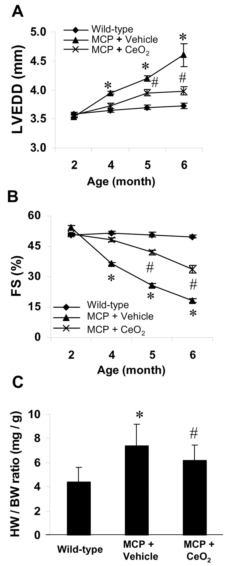
Effects of CeO2 nanoparticles on left ventricular function and remodeling. LV end-diastolic dimensions (LVEDD) (A) and percent fractional shortening (%FS) (B) were depicted from 2 to 6 months of age. The progressive LV dilatation and LV dysfunction in the vehicle-treated MCP mice were attenuated by treatment with CeO2 nanoparticles. *P<0.001 versus wild-type controls; #P < 0.05 versus vehicle-treated MCP mice; n=6 per group and per time point. Heart weight to body weight ratio (HW/BW) was depicted at 6 months of age (C). *P<0.001 versus wild-type controls; #P < 0.05 versus vehicle-treated MCP mice; n=5 per group.