
Fig. 3.
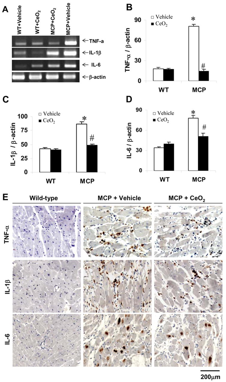
Effects of CeO2 nanoparticles on TNF-α, IL-1β, and IL-6 gene expression in the myocardium of MCP mice. (A) Expression of TNF-α, IL-1β, and IL-6 mRNA in the myocardium of wild-type control, vehicle- and CeO2-treated MCP mice were assayed by RT-PCR. (B–D) Bands were quantified by densitometric analysis and normalized by β-actin. *P < 0.001 versus wild-type controls; #P<0.05 versus vehicle-treated MCP mice; n=5 per group. (E) Representative photomicrographs of in site immunohistochemical staining demonstrating production of proinflammation cytokines TNF-α, IL-1β, and IL-6 in the myocardium. Positive-stained cells were visualized with diaminobenzidine (brown).