
Fig. 5.
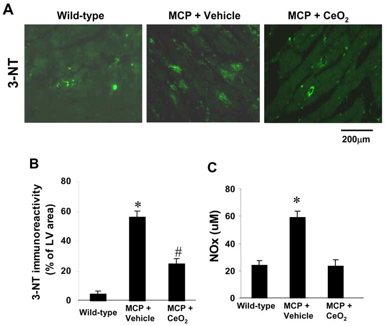
Effects of CeO2 nanoparticles on myocardial oxidative stress. (A) Representativeimmunohistochemical photomicrographs of 3-NT in the myocardium of wild-type control, vehicle-, and CeO2-treated MCP mice. (B) Histograms showing the accumulation of nitrotyrosine formation in the myocardium of different group of mice. *P < 0.001 versus wild-type controls; #P<0.05 versus vehicle-treated MCP mice and wild-type controls; n=5 per group. (C) Serum levels of NOx (total nitrate and nitrite in proteins) assayed by Griess reagent. *P < 0.001 versus wild-type controls and CeO2-treated MCP mice; n=6 per group.