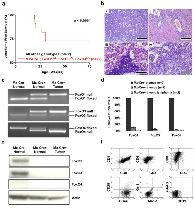
Figure 1. Thymic lymphomas in mice following somatic deletion of three FoxO genes.

a, Thymic lymphoma-free survival of pI-pC treated Mx-Cre+ mice and controls representing combined genotypes including: Mx-Cre+;FoxO1L/L (n=11), Mx-Cre+;FoxO1/3L/L (n=14), Mx-Cre+;FoxO1/4L/L (n=11), and all Mx-Cre− controls (n=36). No lymphomas were observed in controls up to 100 weeks of age. b, Histology and tissue infiltration of thymic lymphoma in Mx-Cre+ mouse, H&E stains. (i) thymus (ii) liver, (iii) lung, and (iv) bone marrow. Scale bars: 200 μm (i and ii) and 100 μm (iii and iv). c, FoxO1, FoxO3, and FoxO4 gene deletions in thymic lymphomas and control thymi by PCR analysis. d, Reduction of mRNA levels of FoxO1, FoxO3, and FoxO4 in Mx-Cre+ endothelial cells. Quantitative real-time PCR performed on Mx-Cre− thymi (7 weeks), Mx-Cre+ thymi (7 weeks), and Mx-Cre+ thymic lymphomas (n=2, 19 and 30 weeks post pI-pC); relative reduction of FoxO levels relative to Mx-Cre− thymi is shown. e, Western blot analysis of Mx-Cre− and Mx-Cre+ thymi and thymic lymphoma samples. f, Flow cytometric analysis of representative thymic lymphoma.
