
Figure 2. Systemic hemangiomas in mice following somatic deletion of three FoxO alleles.
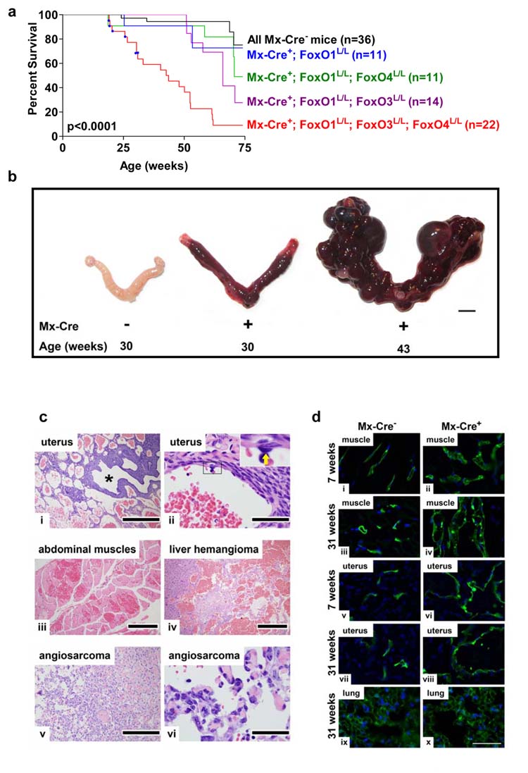
a, Survival of Mx-Cre+ and Mx-Cre− littermates controls. Blue squares indicate deaths due to thymic lymphomas. P value indicates comparison between Mx-Cre+ mice (red) and all Mx-Cre− controls (black). b, Age-dependent progression of uterine vascular lesions in Mx-Cre+ females, scale Bar=5 mm. c, Histology of systemic vascular lesions, H&E stains. Genotypes are Mx-Cre+ unless otherwise noted. i, uterus, low magnification (asterisk: uterine lumen); ii, uterine hemangioma (inset: benign endothelium of hemangioma vascular channels; arrow: endothelial cell); iii, abdominal muscle hemangioma; iv, liver hemangioma, Mx-Cre+ mouse; v-vi, angiosarcoma, Mx-Cre+ mouse. Scale bars: 500 μm (i and v), 400 μm (iv), 200 μm (iii), and 50 μm (ii and vi). d, Fluorescence micrographs of abdominal muscle, uterine horn, and lung endothelium after vascular perfusion of fluorescein-labeled lectin (blue: DAPI labelled nuclei). Note increased endothelial cell density by 7 weeks in muscle and uterine horn but not in lung as late as 31 weeks. Scale bar=50 μm.