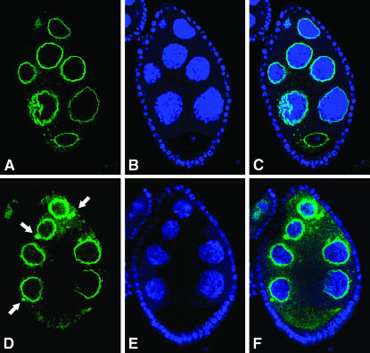
Figure 1.—
GFP-Nup154 and Nup154 protein distribution in transgenic lines. (A–C) Single confocal section of a stage 8 egg chamber expressing one copy of the gfp-nup154 transgene in the germline. (A) GFP-Nup154 is correctly localized at the edge of the nurse-cell nuclei, highlighted in B by TOTO-3 staining. (C) The merged image shows that GFP-Nup154 also decorates the rim of the oocyte nucleus, where the tightly condensed chromosomes occupy a central position. (D–F) Single confocal section of a stage 8 egg chamber expressing two copies of the nup154 transgene in the germline. (D) Nup154 is localized as expected at the nuclear periphery, but an additional signal is detected in cytoplasmic particles localized in the proximity of the nurse-cell nuclei (arrows). (E) TOTO-3 staining. (F) Merged image.

