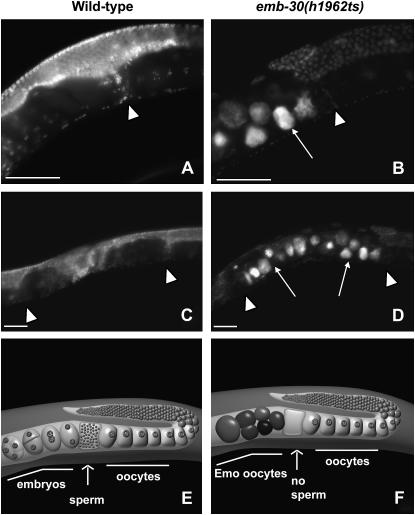
Figure 5.—
The emb-30(h1962ts) allele has an Emo phenotype at 25°. Animals were shifted in L4 stage from 20° to 25° and their adult progeny were DAPI stained as young adults. Images show partial gonad and partial uterus (A and B) or the entire uterus (C and D). In wild-type hermaphrodites (A and C) the highly condensed haploid sperm nuclei can be seen in the spermatheca (arrowhead). Left of the spermatheca, embryos are located within the uterus (A and C). In emb-30(h1962ts) animals (B and D), there is no sperm DNA detected in the spermatheca (arrowhead) and the uterus is filled with Emo oocytes only (arrows). (E) Schematic of a single wild-type adult hermaphrodite gonad arm including half of the uterus. A maturing oocyte is pushed into the spermatheca, successfully fertilized by sperm, and pushed into the uterus where it develops into an embryo. (F) Schematic of a single emb-30(h1962ts) adult hermaphrodite gonad arm including half of the uterus. A maturing oocyte is pushed into the spermatheca. The lack of sperm results in a failure to fertilize. The unfertilized oocytes pass through spermatheca into the uterus, continue cycling, and become Emo, highly polyploid, round, brown oocytes that lack an eggshell. Bars, ∼50 μm.

