
Figure 6.
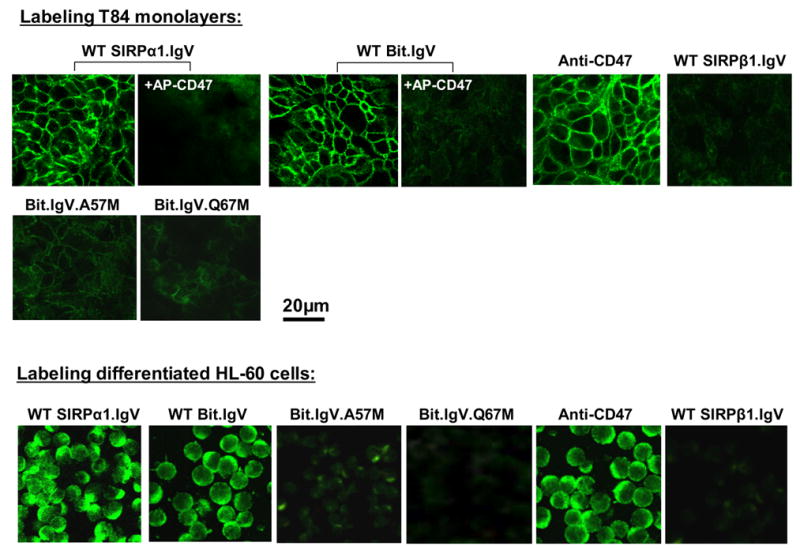
Cell surface labeling epithelial monolayers and HL-60 leukocytes by SIPRα.IgV-Fc fusion proteins. Upper panel: T84 epithelial monolayers were incubated with wild-type or mutant SIRP IgV fusion proteins as labeled in the figure, in the presence or absence of CD47-AP (5μg/ml), followed by detection using fluorescence-conjugated secondary antibody. For control staining, the monolayers were also labeled with anti-CD47 mAb C5D5 (10μg/ml), SIRPβ1.IgV-Fc or Fc only (not shown). Lower panel: similar labeling procedures were also performed to analyze wild-type and mutant SIRPα.IgV-Fc binding to differentiated HL-60 cell surfaces. Images shown represent one of at least three individual experiments, with multiple images taken per slide.