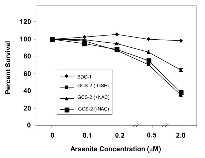
Figure 1.

Determination of the cytotoxicity of arsenite in GCS-2 cells. Dose response to arsenite-induced cytotoxicity in GCS-2 and BDC-1 cells. Cytotoxicity was measured using the MTT assay as described in “Methods”. A fixed number of GCS-2 cells were allowed to grow in 24-well plates in the presence of 2.5 mM GSH or 2 mM NAC for 48 h. GSH was withdrawn for 24 h and both BDC-1 and GCS-2 cells were treated with varying concentrations of arsenite for 21 h. NAC-dependent cells were divided into two groups: One group was grown for an additional 24 h in NAC and treated with different concentrations of arsenite in the presence of NAC while NAC was withdrawn from the other group for 24 h and the cells were treated with varying concentrations of arsenite for 21 h, followed by the addition of 40 μl of MTT reagent to each well. The plates were incubated for 4 h at 37°C and read on a plate reader at 570 nm. Results are presented as mean ± SEM.
