
Figure 5.
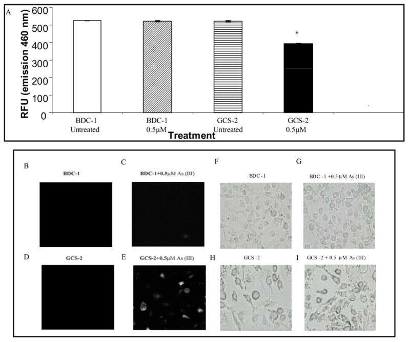
Effect of arsenite on proteasome function in GCS-2 cells. (A) In vitro functional assay for proteasome activity. The chymotrypsin-like peptidase activity of the proteasome was assayed with fluorogenic substrate succinyl-leu-leu-val-tyr-AMC. Cells were treated with arsenite, washed, lysed, and assayed for chymotrypsin activity. Values are presented as mean ± SEM for 5 determinations. p ≤ 0.001; RFU, relative fluorescence unit. (B) In vivo functional assay for the proteasome. BDC-1 and GCS-2 cells were transfected with pGFPu containing neomycin as described under “Materials and Methods”. Stably transfected cells were isolated, treated with and without arsenite and 21 h later the cells were assayed for intracellular fluorescence by confocal microscopy. B, D, F, and H are untreated cells; C, E, G, and I are cells treated with 0.5 μM arsenite. B-E is confocal micrographs of GFPu expression in cells. F-H is the corresponding light micrographs.