
Figure 1.
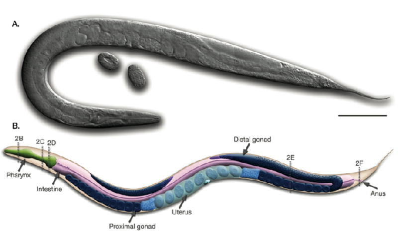
C. elegans hermaphrodite, left lateral view. (A) Micrograph of a hermaphrodite using differential interference optics. Two oval-shaped embryos can be seen near the vulval opening of the animal. (B) Diagram of the major anatomical features of the hermaphrodite. At the anterior (on the left) of the animal the distal gonad bends behind the intestine whereas at the posterior (on the right) the entire U-shaped gonad is visible (shown in shades of blue). The dotted lines labeled 2B–F correspond with the cross sections B–F in Figure 3. Scale bar represents 0.1 mm. Reprinted from Wormatlas (www.wormatlas.org) with permission.