
Figure 1.
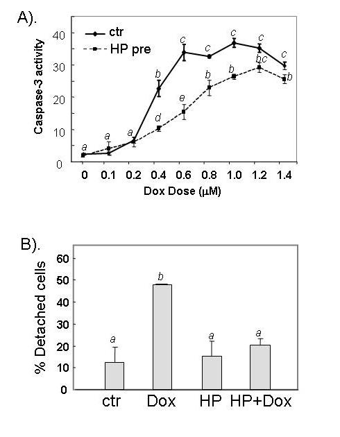
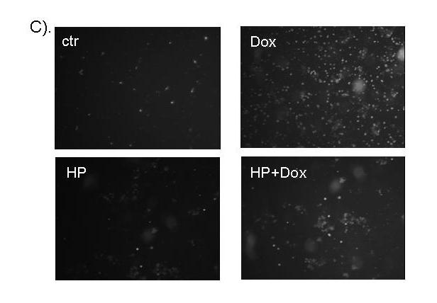
H2O2 pretreatment reduces Dox-induced apoptosis. Serum-starved CMCs were treated with 100 μM H2O2 for 1 hr. The cells recovered for 24 hr in fresh DMEM containing 0.5 % FBS before treatment with different doses of Dox (A) or 0.6 μM Dox (B & C). Caspase-3 activity (A), cell detachment (B) and Annexin V binding (C) were measured 16 hr after addition of Dox. The relative fluorescent unit (RFU) was corrected for protein content to indicate caspase-3 activity (A). At least 300 cells were scored from each view under a phase contrast microscope and three views were chosen randomly for scoring the proportion of detached versus attached cells (B). A letter indicates a significant difference (p<0.05) from the means labeled with a different letter as determined by ANOVA followed by Bonferroni analysis (A, B).