
Figure 3. Inhibition of human glioma xenograft growth in vivo by interference with HH-GLI signaling.
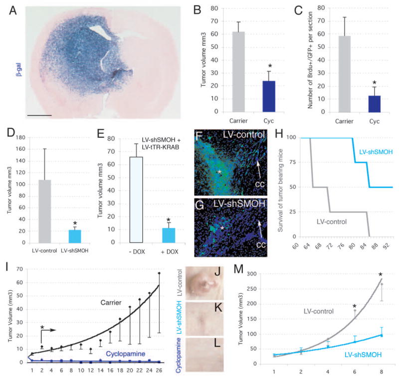
A) Development of invasive gliomas after intracerebral implantation of gliomaspheres transduced with a lentiviral vector expressing LacZ ±1 month after implantation of 105 cells (n=7 mice for GBM-8; n=7 mice for GBM-7 and n=7 mice for OG-III-1, not shown). Glioma stem cell-derived tumor cells infiltrate into the parenchyma (cells are blue after X-Gal cytochemistry).
B,C) Inhibition of intracraneal tumor growth after treatment with cyclopamine (n=3 mice for OG-III-1, n=3 mice for GBM-8, not shown) but not carrier alone (n=3 mice for OG-III-1, n=3 mice for GBM-8, not shown). Tumor volume (B) or BrdU+ cell number (C) were monitored in section reconstructions.
D) Tumor volume was also decreased after LV-shSMOH transduction (n=3 mice) but not after LV-control transduction (n=3 mice).
E) Infection with GBM-8 gliomasphere cells with LV-shSMOH and LV-tTR-KRAB (see supplemental information) reduced tumor volume only after induction by DOX (n=3 mice with DOX treatment and 3 mice without).
F,G) Representative examples of intracraneal gliomas ±1 month after implantation of 104 GFP+ gliomapsheres treated as described. The asterisk denotes the injection site. Note the strong migration of tumor cells contralaterally along the corpus callosum (cc, arrow) in control (G) but not LV-shSMOH-expressing cases (G).
H) Survival of mice harboring LV-control (n=4) or LV-shSMOH-transduced (n=4) cells. Mice were sacrificed at the first signs of terminal disease before extreme pain was apparent.
I-M). Inhibition of subcutaneous U87 gliomas by injection of cyclopamine (I, L; n=8 tumors) or interference with SMOH (LV-shSMOH; J,K,M; n=6 tumors), but not in U87 tumors treated with carrier alone (I and not shown; n=8 tumors) or infected with LV-control (J,M; n=6 tumors). Asterisks denote significative changes (p<0.05).
Scale bar= 0.25cm (A), 0.8mm (F,G) and 8mm for (J–L).