
Figure 2. Blocking wnt signaling with sFz5 reduces T cell transmigration.
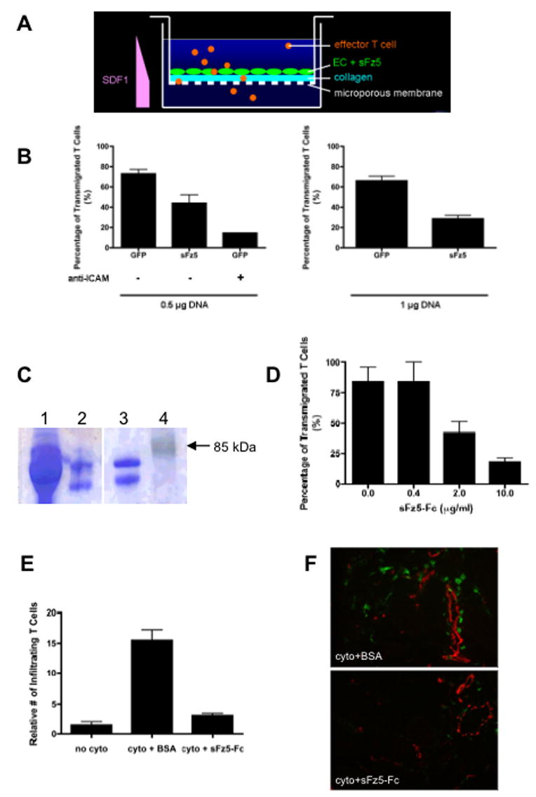
(A) Model system for T cell transmigration assays. (B) EC were transfected with 0.5 μg of GFP or sFz5 (left) or 1 μg of GFP or sFz5 (right), grown to confluence on collagen I-coated inserts, and then stimulated with TNF-α (10ng/ml) for 4hr to induce expression of adhesion molecules. Effector T cells were plated in the upper well in the presence of HB64 (−) or anti-ICAM-1 mAb (left). SDF1-α (100ng/ml) was added to the lower well. Cells that transmigrated into the lower well after 24 hours were counted. Results are representative of more than three independent experiments. Mean and SD of triplicate wells, * – p<0.05 (t-test) for GFP v sFz5, and p<0.005 (t-test) for control, v anti-ICAM-1 mAb. (C) Coomassie blue staining of an SDS-PAGE gel showing purified sFz5-Fc produced in HEK293 cells (lane 2), or purchased (lane 3). Supernatant from transfected HEK293 cells prior to purification is shown in lane 1. MW markers are in lane 4. (D) Effector T cells were added to untransfected EC monolayers in the presence of 10μg/ml BSA or sFz5-Fc protein. Transmigrated T cells were counted after 24 hours. Mean and SD of triplicate wells, * – p<0.005 (t-test) for control (0μg/ml sFz5-Fc, 10μg/ml BSA) v 10μg/ml sFz5-Fc. One of three similar experiments. (E) Mice were injected i.d. on the back with TNFα (10ng) and IFNγ (300U), or with TNF + IFNγ along with BSA or sFz5-Fc (1μg each). Controls received vehicle (PBS) alone, and all injections were in a total volume of 10μl. Skin was harvested after 22 hours and frozen. Sections were stained for CD3 (green) to show T cells and CD31 (red) to highlight blood vessels. Six fields from duplicate sites from each mouse were analyzed (blinded) for each condition. Mean and SD, * – p<0.01 (t-test) for BSA+cytokine v sFz5-Fc+cytokine. One of five similar experiments. (F) Representative sections stained for T cell CD3 (green) and EC CD31 (red). Conditions as indicated.