
Fig. 2.
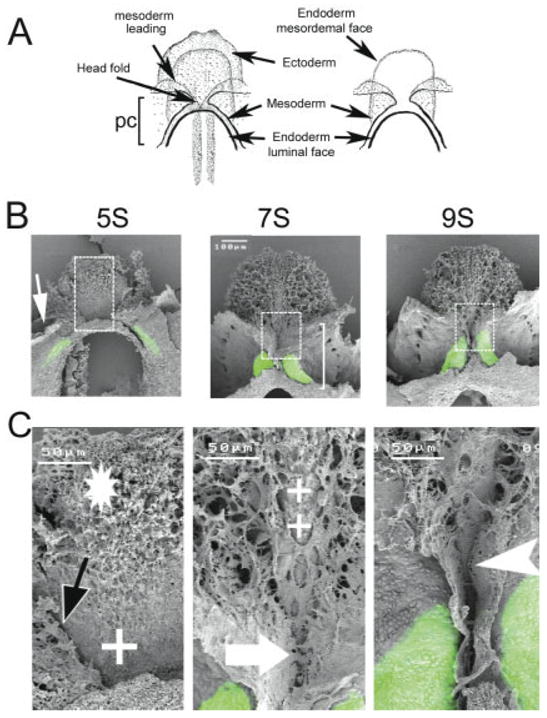
Fusion of the heart-forming regions. Scanning laser confocal micrographs were obtained from embryos (14) immunostained with MF20 to show sarcomeric myosin localization. The head ectoderm was removed from each embryo prior to being processed for scanning electron microscopy. A: Representation of an embryo before (left) and after (right) ectoderm removal (ventral view). The myosin expression pattern (green) was overlaid onto the sister SEM micrograph for each embryo. B: SEM/MF20 overlays from embryos at pre-fusion (5-somite stage), fusion (7-somite stage), and the post-fusion period (9-somite stage) are shown. C: Higher magnification of boxed region shown in B. At the pre-fusion stage (5 somites), the coelom is beginning to form (white arrow). Note that the MF20 localization is lateral with respect to the leading edge of the mesoderm layer (black arrow) at this stage of development. The endoderm layer (mesodermal face) has mesenchymal cells in the prechordal plate (*), and more caudally, the endoderm is without mesenchymal cells (+). At the 7-somite stage, sarcomeric myosin is expressed in the splanchnic mesoderm next to the region of the mesoderm fusion. The heart occupies only half of the pericardial coelom (white bracket). Note that the anterior portion of the pericardial coelom (above the large white arrow) has not fused. Some areas of the endoderm also lack mesenchymal cells (++). At the 9-somite stage, myosin expression extends over the length of the primitive heart canal and the ventral fusion line extends more cephalically (compare 7- to 9-somite images in C). In addition, the ventral fusion line extends caudally (white arrowhead). pc, pericardial coelom.