
Fig. 4.
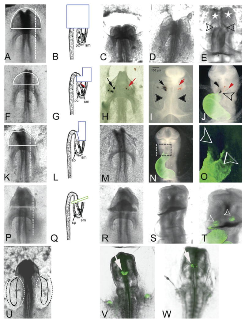
(Overleaf) Ablation mapping of cardiogenic potential of mesoderm from regions anterior to the pericardial region of the coelom in 6-somite stage embryos. Ventral views (A,F,K,P,U) of representative 6-somite embryos and cross-sectional diagrams (B,G,L,Q) of the corresponding surgical procedures performed. The positional relationship of the surgery with respect to the heart-forming regions is highlighted in U (see Fig. 1A). A–E: (n = 1) Although the entire head was removed (t = 0; C) the heart began to form within 3 hr (D). After 24 hr in culture (E), a conus-like structure with two-limb “biconal heart” (open arrows) was observed. The looping of the heart looks abnormal, and the head structures are missing. However, the head mesenchyme was partially restored (white stars). F–J: (n = 5) The ventral ectoderm, mesoderm, and endoderm of the anterior half of the head were removed (solid white line). H: After ablation the most anterior mesoderm tissue was labeled with black ferric-ferrous oxide and red ferric oxide (black and red arrows, respectively). The same embryo after 12 hr of incubation (I) and after 24 hr of incubation (J; MF20 immunostain overlay in green). Note that iron particle labels after 12 hr of culture (I) are outside the heart and far from the ventral middle line. Twenty-four hours later (J), the heart apparently is well developed with coincident distribution of both labels at the distal aspect of the ventral fusion line (open arrow), as well as dispersed individually (black and red arrows) in the lateral regions of head mesenchyme. K–O: (n = 15) Ectoderm alone was removed from the anterior half of the head (solid white line). M: The same embryo immediately following the ablation and (N) again after 24 hr in culture (16 somites; HH12). The organization of the head was not affected. O: Higher magnification of boxed area in N. Note that the outlet has two limbs (open arrows). P–T: (n = 3) An incision was made with a glass needle at the anterior border of the pericardial region of the coelom. Once ectoderm and endoderm were opened, we inserted a small piece of eggshell membrane (green box in Q) and the embryo was allowed to develop 24 hr in culture (S). T: Higher magnification of MF20 immunostained embryo in S. Note the heart was apparently well developed and the primitive outlet was below the eggshell membrane. Only two clusters of cells were MF20-positive above the eggshell membrane (open arrowheads). The growth of the head was not affected. U–W: Both mesoderm and endpderm were removed from posterior bilateral margins of the embryo (dotted line) that included the cardiogenic region as described by Stalsberg and DeHaan (1969). Two independent embryos that developed to the 16-somite stage are shown (V and W). The MF20 staining was present as two distinct masses on the foregut. pc, pericardial region of the coelom; sm, somatic mesoderm; sp, splanchnic mesoderm.