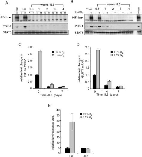
Figure 2.
Growth factors regulate the expression and activity of HIF-1α. (A) Cells cultured in the presence (+IL3) or absence of IL-3 at the indicated time points were incubated in normoxic (N) or hypoxic (H) conditions for 4 h and HIF-1α protein levels were analyzed by Western blot. Cells cultured in the presence of IL-3 and treated with 100 μM CoCl2 were used as a control. (B) At similar time points following withdrawal from IL-3, cells were treated with PBS or 100 μM CoCl2 for 4 h and analyzed by Western blot for HIF-1α and PDK-1 protein levels. Lysates from cells cultured in the presence of IL-3 exposed to hypoxia were used as a control. STAT3 was used as a loading control. Cells were cultured in the presence or absence of IL-3, and at the indicated time points, total RNA was isolated and analyzed for Hif-1α (C) or Glut1 (D) mRNA levels by real-time qPCR. Data are a representative experiment for triplicate samples ± SD. (E) HIF-1α promoter activity in response to hypoxia is lost during growth factor withdrawal. HIF-1α-responsive promoter containing three tandem HRE from the murine pgk1 was transfected into cells cultured in the presence or absence (3 wk) of IL-3. Ten hours after transfection, cells were incubated in normoxic (21% O2) or hypoxic (1.5% O2) conditions for an additional 24 h. Promoter activity is expressed as a ratio of luciferase to Renilla activity (relative luminescence units). Data are a representative experiment ± SD. for triplicate samples.

