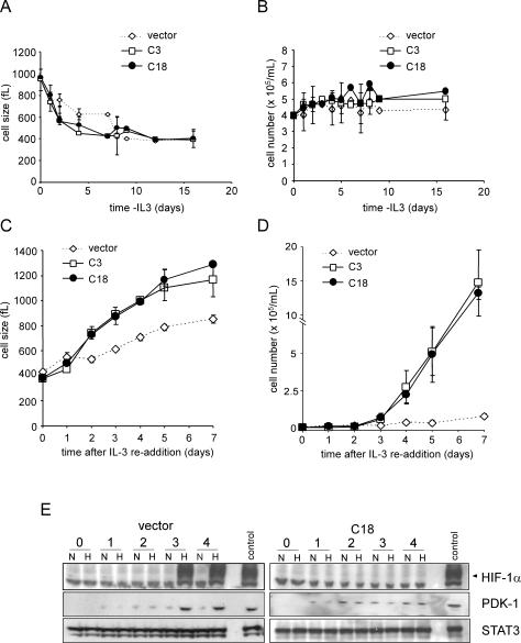
Figure 6.
Cell growth and proliferation are enhanced in the absence of HIF-1α. Cells were withdrawn from IL-3 and size (A) and cell number (B) were measured at the indicated time points. On day 16 of withdrawal, cells were restimulated with IL-3 and size (C) and cell number (D) were measured. Data represent the mean of three independent experiments ± SD. (E) Cells cultured in the absence of IL-3 for 16 d were placed in fresh complete medium containing IL-3. At the various time points, cells were subjected to an additional 4 h of culture under 21% O2 (N) or 1.5% O2 (H). Cell lysates were analyzed by Western blot for HIF-1α and PDK-1 expression. A representative vector control and a HIF-1α cell line (C18) expressing a stable HIF-1α shRNA are shown. STAT3 was used as a loading control. Cells cultured in the presence of IL-3 and exposed to hypoxia were used as a positive control.

