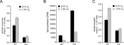
Figure 8.
Glucose utilization in primary T cells is impaired by HIF-1α. (A) Glucose consumption in primary T cells isolated from Hif-1αfl/fl Cre− (wild-type [WT]) or Hif-1fl/fl Cre+ (Cre) mice that were administered tamoxifen. T cells were stimulated under 21% or 1.5% oxygen by plate-bound anti-CD3/anti-CD28 in 1-mL cell cultures, and supernatants were collected 48 h post-stimulation for glucose analysis. Data are a representative experiment performed in triplicate ± SD. (B) Cells were activated by plate-bound anti-CD3/ anti-CD28 for 2 d followed by addition of 14C-labeled pyruvate under normoxic or hypoxic conditions. Cells were cultured for an additional 2 d in the presence of IL-2. Lipids were extracted from cell lysates and total incorporation of 14C-labeled lipid was measured by scintillation counting. Data are a representative experiment ± SD for triplicate samples. (C) Cells were cultured under identical conditions described in A and the amount of secreted lactate into 1 mL of culture medium was measured as described in Materials and Methods. Data are a representative experiment ± SD.

