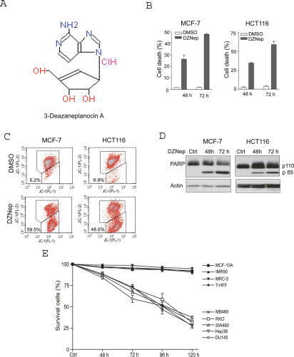
Figure 1.
DZNep preferentially induces apoptosis in cancer cells. (A) Chemical structure of DZNep. (B) MCF-7 and HCT116 cells were treated with 5 μM DZNep for 48 and 72 h, followed by PI staining and FACS analysis. (C) MCF-7 and HCT116 cells were treated with DZNep for 72 h, followed by JC-1 staining and FACS analysis. MTP was quantified by the cells with lower membrane potential (ΔΨm). (D) MCF-7 and HCT16 cells were treated with 5 μM DZNep for 48 and 72 h, and whole-cell extracts were analyzed by Western blotting. Cleavage of PARP was detected after DZNep treatment. β-Actin was used as a loading control. (E) Cell death response of a variety of cancer cells and normal cells to DZNep. Indicated cells were treated with 5 μM DZNep for up to 120 h and the cell death was measured by PI staining and FACS analysis. Data represent ±SD from three independent experiments.

