
Fig. 1.
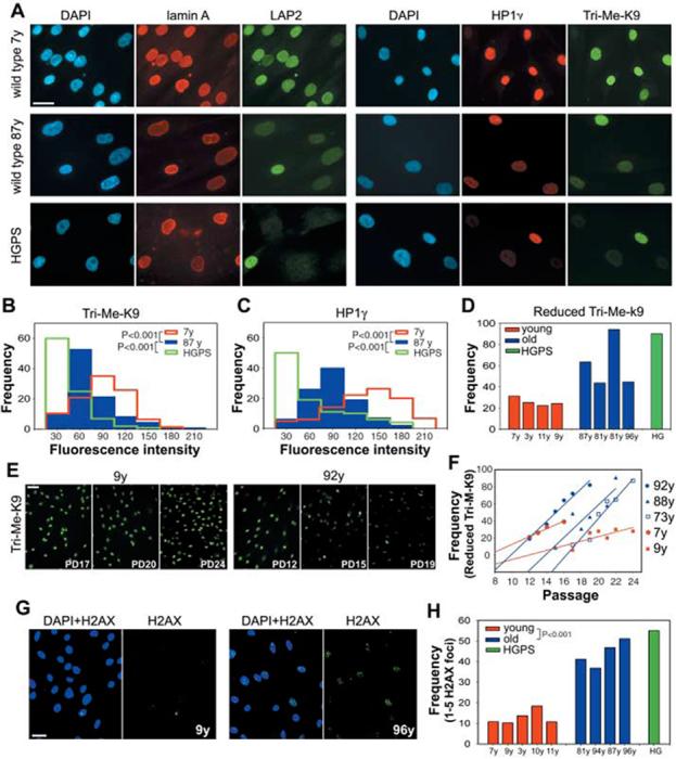
Nuclear abnormalities in cells from old individuals. (A) Immunofluorescence microscopy on primary dermal fibroblasts from young (7 y) and old (87 y) healthy individuals and a HGPS patient. Scale bar, 10 μm. DAPI, 4′,6′-diamidino-2-phenylindole. Intensity distributions of the average fluorescent signal for (B) Tri-Me-K9H3 and (C)HP1γ in fibroblasts from healthy individuals of indicated age and a HGPS patient. (D) Quantitation of cells showing reduced amounts of Tri-Me-K9 in passage-matched population doublings (PD) 19 cell lines from healthy individuals of indicated age and a HGPS patient (9). (E and F) Reduction of Tri-Me-K9 H3 over cell passage. Best linear fits are shown. (G and H)Increased DNA damage in passage-matched (PD20) cell lines from healthy individuals of indicated age and a HGPS patient detected by antibody to H2AX. Scale bar, 20 μm. N > 200.