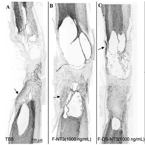
Figure 1.

A-C. Neuronal fiber (Tuj1) staining of sagittal sections of lesion areas treated with (A) TBS, (B) F-NT3 (1000 ng/mL), and (C) F-DS-NT3(1000 ng/mL) after 12 weeks. Substantial cavitation and expansion of the lesion site occurred in all groups. Parasagittal sections, with rostral cord oriented toward top, ventral surface toward right. Encroaching dorsal and ventral roots (arrows).
