
Figure 3.
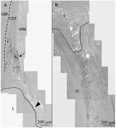
Corticospinal and ascending sensory tract tracing at 12 weeks. Parasagittal sections, with rostral cord oriented toward top, ventral surface toward left. A. Biotin dextran amine (BDA) anterograde tracing of the corticospinal tract (CST) in a F-DS-NT3 (1000 ng/mL)-treated cord. The CST extends from the rostral intact cord toward the lesion site. The CST is located just dorsal to the gray matter (GM, indicated by dotted line), in the most ventral part in the white matter (WM) of the dorsal funiculus. In all groups, as fibers approached the lesion (L, border indicated by line), the fibers were truncated or diverted to more dorsal white matter (arrow). Infrequently, CST-positive fibers were seen in the spared dorsal matter encircling the rostral lesion cavity (arrowhead). B. Tracing of ascending sensory neurons with cholera toxin B (CTB) in a n F-DS-NT3(1000 ng/mL) treated cord. In all groups, CTB staining ended abruptly at the lesion border with the intact cord (IC).