
Fig. 1.
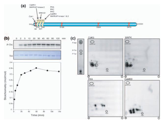
Phosphorylation of TH by Cdk5 and phosphoamino acid and phosphopeptide map analysis. (a) Schematic diagram of TH with sites of in situ phosphorylation indicated (yellow) along with protein kinases. Positions of all serine residues followed by proline are indicated in red and labeled, except for Ser31. (b) Time-course of in vitro phosphorylation reaction. Radiolabeled (P-TH) and Coomassie stained (TH) TH phosphorylated by Cdk5 and quantified stoichiometry (bottom). (c) Phosphoamino acid analysis of 32P-labeled TH phosphorylated by Cdk5 is shown, top left. The positions of comigrating phosphoserine, phosphothreonine and phosphotyrosine standards and the origin are indicated. Tryptic phosphopeptide maps of TH phosphorylated in vitro with the indicated protein kinases are shown in the four larger panels. The positions of comigrating phosphopeptides are indicated by numbers. The positions of the origins and phenol red markers are also indicated.